المقالات
Peripheral Ossifying Fibroma

Peripheral ossifying fibromas are well-defined oral growths that contain bone tissue. They are most often found attached to the gums near the molars or premolars, and are usually connected either by a wide base of tissue or by a stalk-like growth. Usually, they are pink in color, much like the color of the gum tissue. As they enlarge, they may become irritated and more red in color. These lesions are commonly associated with poor oral hygiene and early periodontal (gum) disease, also called gingivitis.
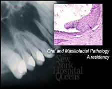
Peripheral ossifying fibromas are smooth and firm to quite hard, depending on the amount of bone they contain. X-ray examination of these lesions will typically show the central mass of bone. They normally do not bleed easily, but often become ulcerated and reddened due to an infection that is caused by irritation from chewing or vigorous toothbrushing.
These lesions can occur at any age, but are most common in children and young adults, and occur twice as often among females. They can be found in the upper or lower jaw, usually behind the molars.
We will remove the fibroma surgically and correct any source of irritation in the area. Healing in the mouth is normally fairly rapid and
usually occurs without any problems, but these lesions have been known to recur (re-develop) occasionally. Without treatment, they can become very large and interfere with normal chewing and swallowing. On occasion, very large growths may cause obvious swelling or enlargement in the face.
