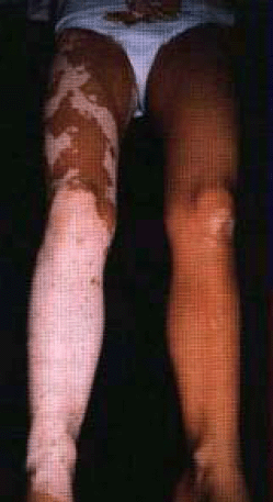
Vitiligo البهاق (فقد الصباغ الجلدي- فقد الميلانين)
INSTRUCTION
Look at this patient.
SALIENT FEATURES
History
-
Ask whether or not the disorder runs in the family (familial in 36% of cases).
-
Other autoimmune disorders (hypothyroidism, hyperthyroidism, thyroiditis,Addison's disease, diabetes mellitus, pernicious

Examination
-
Hypopigmented patches which are distributed symmetrically:some-times the border may be hyperpigmented. The distribution often includes wrists,axillae, perioral, periorbital and anogenital skin.
-
White hairs in the vitiliginous area.
-
Some spontaneous repigmentation in sun-exposed regions (in a third of cases).
Proceed as follows:
Look at the scalp for alopecia and white hair.Note. Scratching when the disease is active may induce lesions along the scratchmarks; this is termed isomorphic response or Koebner's phenomenon.
DIAGNOSIS
This patient has vitiligo (lesion) which is of autoimmune origin (aetiology) and can becosmetically distressing to the patient (functional status).
ADVANCED-LEVEL QUESTIONS
Mention some associated conditions.Organ-specific autoimmune conditions:
-
Thyroid disease - Graves' disease, myxoedema, Hashimoto's disease.
-
Pernicious anaemia.
-
Diabetes mellitus.
-
Alopecia areata.
-
Addison's disease.
Which fungal condition can be mistaken for vitiligo?
Pityriasis versicolor (caused by the fungus Malasseziafurfur).
How would you manage such patients?
Cosmetics are useful for concealing disfiguring patches.
When less than 20% of the skin is involved, topical methoxsalen withlong-wavelength ultraviolet A (UVA) is used, followed by thorough washing andapplication of an SPF 15 sunscreen.
When more than 20% of the skin is affected, oral methoxsalen with UVA.
When vitiligo is extensive, good cosmetic results may be obtained by'de-pigmenting' normal skin with a bleaching agent like hydroquinone.
New lesions of vitiligo may benefit from topical steroids.
Newer therapies include epidermal autografts and cultured epidermis combinedwith PUVA.Note. On the whole, treatment of vitiligo remains unsatisfactory.
Mention a few conditions in which hypopigmentation is common
Hypopituitarism.
Albinism.
Phenylketonuria.
Leprosy.
Burns.
Radiodermatitis.
Piebaldism (an autosomal dominant condition manifested by a white forelock).
Ash leaf spots (tuberous sclerosis).
Leukoderma (a disorder that occurs as a complicaton of lichen planus, lichensimplex chronicus, atopic dermatitis and discoid lupus erythematosus).
What is the histology of vitiligo?
Characteristically, there is partial or complete loss of pigment-producing melanocytes inthe epidermis. In contrast, some forms of albinism have melanocytes but no melaninpigment is produced because of lack of, or a defect in, the tyrosinase enzyme.
Why are melanocytes progressively lost in this condition?
The various theories of pathogenesis include:· Autoimmune destruction due to circulating antibodies against the melanocytesand impaired cell-mediated immunity.· Self-destruction by toxic intermediates of melanin production.· Neurohumoral factors.
